
Optos Optomap
"Your eyes are the window to your health"
Optomap Ultra-Widefield Retinal Imaging
The optomap is a wide field retinal image that we offer as part of our full comprehensive eye examination. A thorough evaluation of the back of the eye is critical component of your yearly check up and offers valuable insight on the health of your eyes.
Book Online
Optomap Imaging
the retina
The retina is a nerve layer located in the back of the eye and works as a light organ that detects incoming light and sends feedback to the brain. Early signs of various systemic diseases manifest in the retina before patients experience any subjective signs and symptoms. Some systemic diseases that affect the retina are diabetes, high blood pressure, high cholesterol, colon cancer, and many others!
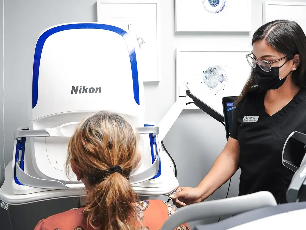
digital retinal imaging
The optomap is a non-invasive, diagnostic tool that produces high resolution images of your retina, optic nerve, and other retinal structures at the back of your eye. These retinal images facilitate early detection of ocular diseases, such as glaucoma, diabetic retinopathy, retinal detachment, holes or tears in the retina, and macular degeneration.
importance of monitoring changes
These wide view, highly detailed, digital retinal images give us an objective data point that allows our doctor to detect and measure changes in the structures of the back of the eye. At the end of your eye examination, we take the time to display your images on a large screen so we can review them together and answer any questions you may have. Our doctor believes in the importance of educating you on your eyes and what steps we need to take to keep them as healthy as possible. These images are compared for changes every year when we see you for your eye examination so we can continue to take care of your eyes.
importance of digital retinal imaging
The optomap allows for early detection and diagnosis of life-threatening diseases like cardiovascular diseases (high blood pressure, diabetes), cancer, and stroke. Early detection can lead to early treatment which can reduce the risk of damage to important structures in the back of the eye which could cause vision loss.
Video
optomap diagnostic tool
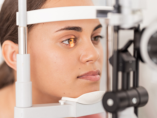
Our exam not only consist of giving you the clearest vision, our doctor also needs to look at the back of your eye, the retina, to check that it is healthy and not showing any signs of disease.
optomap retinal exam
Having a comprehensive eye exam with a retinal image is crucial tool to monitor your eye health and protecting your and your family's eyesight.
Frequently Asked Questions
Why is a retinal exam so important?
Some of the first signs of diseases such as stroke, diabetes and even some cancers can be seen in your retina, often before you have other symptoms. An optomap makes it easier to diagnose and monitor these conditions.
What is an optomap?
The optomap is a digital image of the retina produced by Optos scanning laser technology. It is the only technology that can capture 82% view of your retina at one time. An optomap image does not replace dilation and we sometimes perform both together.
How will optomap benefit me?
The ultra-widefield optomap may help your eye doctor detect problems more quickly and easily. Unlike traditional retinal exams, the optomap image can be saved for future comparisons. We recommend that we repeat the optos image at each routine eye exam.
Are there side effects?
The optomap images are created by non-invasive, low-intensity scanning lasers. No adverse health effects have been reported in over 150 million sessions. It has been said by patients that the bright light mimics the light of Thor.
Is an optomap safe for children?
Yes, in fact, many vision problems begin in early childhood, so it's important for children to receive quality routine eye care.
Does it hurt?
No, it is completely comfortable and the scan takes less than a second. Patients have only experience a bright light.
How often should I have an optomap?
We recommended that you have an optomap each time you have an eye exam.

Contact Info
Office Hours
- Monday 9:00 am - 6:00 pm
- Tuesday 9:00 am - 6:00 pm
- Wednesday 8:30 am - 5:00 pm
- Thursday 8:30 am - 5:00 pm
- Friday 8:30 am - 5:00 pm
- Saturday 8:15 am - 1:00 pm
- (2nd and 4th Saturdays in the month only)
- Sunday Closed
© 2026 Covina Hills Optometric Group. All rights Reserved. Accessibility Statement - Privacy Policy - Sitemap
Powered by: